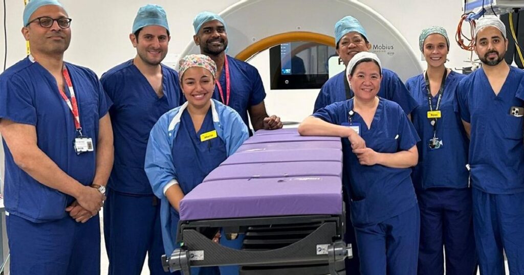
The deep brain stimulation procedure at Queen’s Hospital — mainly for patients with movement disorders like Parkinson’s — needs the brain to be scanned before and after surgery to make sure the electrodes are placed correctly.
But Barking, Havering and Redbridge University Hospitals NHS Trust (BHRUT), which runs Queen’s, said this meant patients having to be moved from the operating theatre to the hospital’s radiology department on a different floor and back again twice during the procedure.
Now neurosurgeon Abteen Mostofi is using a mobile CT scanner during the operation itself, saving up to 90 minutes.
“Moving a bed with a ventilator and pumps through a corridor to the lift takes a lot of logistics and time,” he said.
“But the mobile scanner is much quicker and therefore safer.”
It had taken most of the day for a major operation, according to BHRUT.
But now surgeons can finish in half the time which frees up theatres for more operations.
Stanley Smith, a 67-year-old grandfather who was diagnosed with Parkinson’s in 2019, was one of the first patients at Queen’s to benefit.
He said: “It was a good idea not having to whip you out and run you up the corridor only to come back again.
“My consultant spoke about the deep brain implants and I thought I’d give it a go. It’s a few weeks on from the operation and I’m doing well.”
His quality of life had been “non-existent” for several years due to body tremor and barely being able walk.
Now Stanley works with his neurologist using his pacemaker to stimulate the electrodes in the brain to reduce symptoms.
The pacemaker designed specifically for neuro-surgery works as a GPS scanner for the brain, making sure all the probes are in the right place.
The pacemaker is used to stimulate the electrodes.
It is an extremely delicate manoeuvre implanting electrodes in very small parts of the brain — surgeons have to know exactly where everything is.


